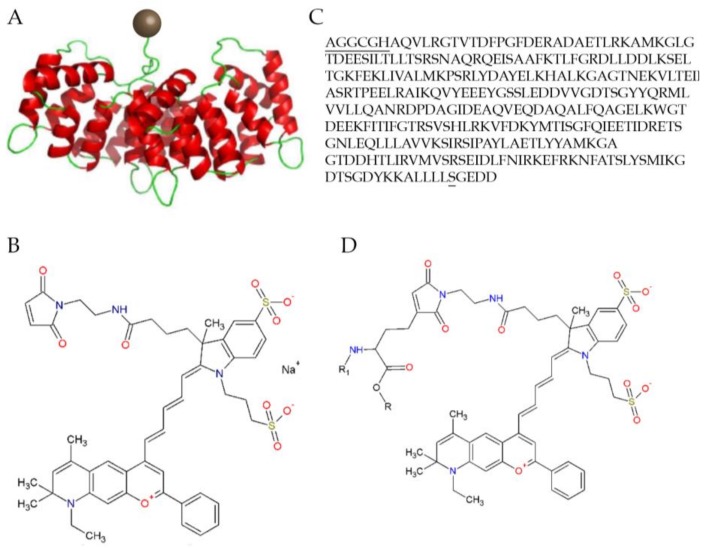
Figure 3.
The structure of the Dy-776-mal labelled annexin V128 molecule: (A) Diagrammatic representation with the Dy-776-mal label represented with a brown sphere and the annexin molecule in red and green; (B) The molecular structure; (C) The optimized amino acid sequence of Anx V128, with the additional amino acids at the N-terminus containing the cysteine and mutated serine; and (D) The molecular structure of the fluorescent conjugation of ANX776 [85].

