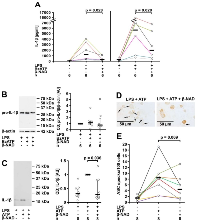
Figure 2.
β-NAD inhibits ATP-induced IL-1β release by primary peripheral blood mononuclear leukocytes (PBMCs). (A–C) PBMCs from healthy donors were left untreated or pulsed with LPS (5 ng/mL) during the process of leukocyte isolation, cultured for 3 h, and stimulated with BzATP (100 µM, 30 min) in the presence or absence of β-NAD (1 mM). (A) The concentration of IL-1β was measured in the cell culture supernatant by ELISA. (B,C) Western blot analysis of cell lysates or concentrated cell culture supernatants using antibodies that recognize pro-IL-1β and mature IL-1β. (B) Representative Western blot of cell lysates; pro-IL-1β is detected with an apparent molecular mass of about 34 kDa. A faint signal corresponding to mature IL-1β was obtained in lysates of cells treated with BzATP and β-NAD only in one out of 6 blots. β-actin (40 kDa) was detected on the same blots as a loading control. (C) Representative Western blot of cell culture supernatants (one out of 8); only mature IL-1β is detected with an apparent molecular mass of 17 kDa. The optical density (OD) of the immuno-positive bands was measured and the values of the samples from cells stimulated with LPS and BzATP were set to one arbitrary unit (AU). Data are presented as individual data points, bars indicate median, whiskers encompass the 25th to 75th percentile. (D,E) LPS-pulsed PBMCs were stimulated with ATP (1 mM) and again, β-NAD (1 mM) was added in some experiments. (D) ASC immunoreactivity in adherent PBMCs was detected in brown color by immunocytochemistry; arrows are pointing to ASC specks. (E) The number of ASC specks per 100 PBMCs was quantified. Data points from individual blood donors are connected by lines in different colors, bars indicate median (A,E); Wilcoxon signed-rank test.

