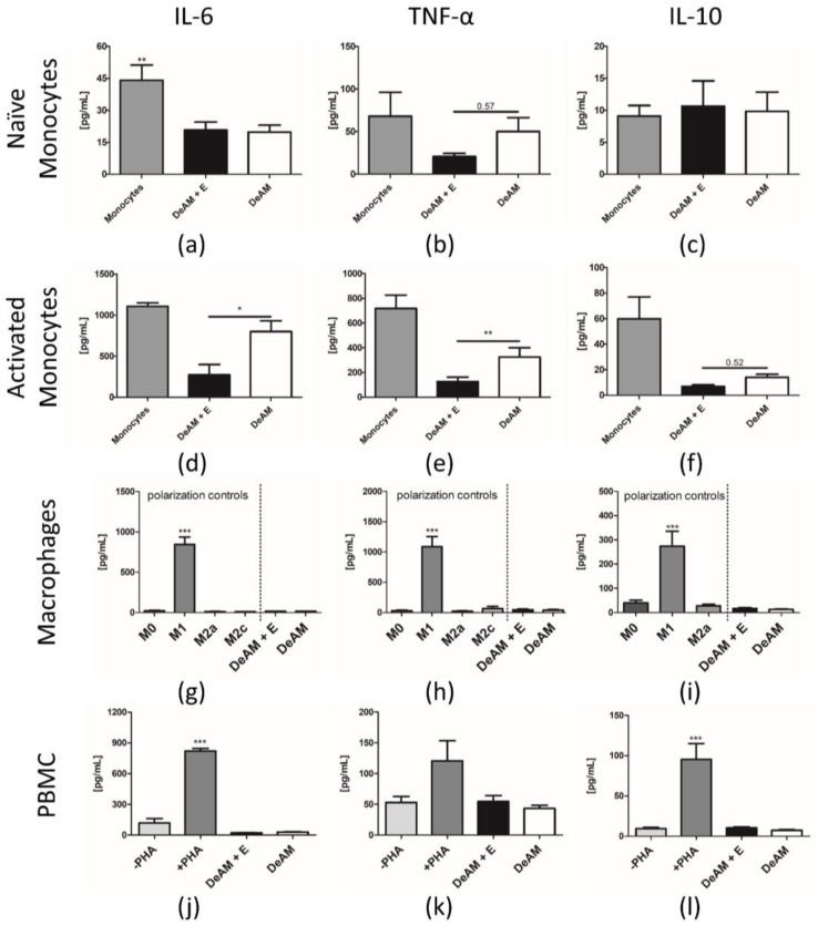
Figure 5.
Cytokine release from monocytes, macrophages and PBMCs cultured on DeAM and DeAM + E scaffolds determined by ELISA. Supernatants were collected after 24 h of naïve ((a–c) ** p < 0.01 to all groups) and LPS-activated ((d–f) * p < 0.05, ** p < 0.01) CD14+ monocytes on DeAM + E (black), DeAM (white) or monocyte standard culture control conditions (grey). Macrophages derived from CD14+-monocytes (M0) were polarized towards pro-inflammatory M1- and anti-inflammatory M2a- and M2c-type. After 2 days, supernatants were collected and analyzed for IL-6, TNF-α and IL-10 ((g–i) *** p < 0.001 to all groups) concentration. PBMCs from human buffy coat were cultured for 5 days. Supernatants were collected and analyzed for cytokine secretion of IL-6, TNF-α and IL-10 ((j–l) *** p < 0.001 to all groups). The positive control group was stimulated with PHA. n ≥ 3.

