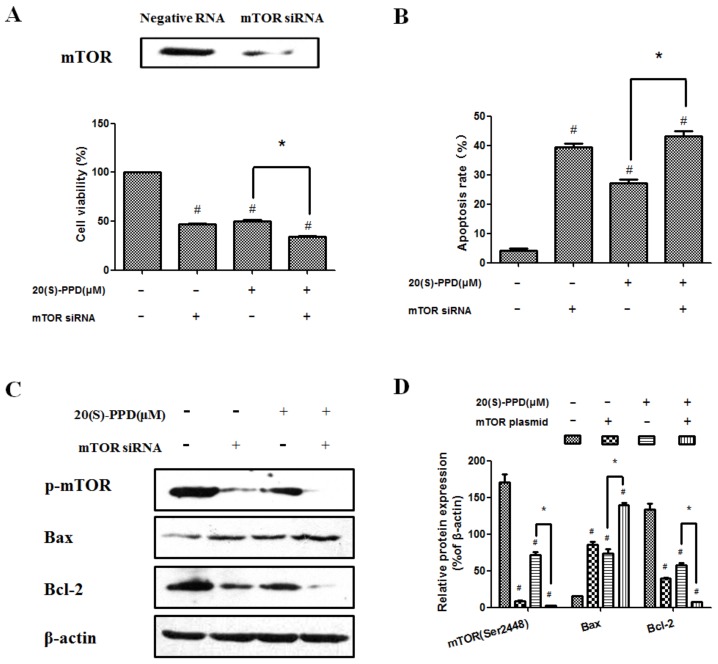
Figure 5.
20(S)-PPD-induced apoptosis was promoted by knockdown of mTOR with siRNA. (A) Western blot was used to detect mTOR expression after siRNA transfection (upper line). After 20(S)-PPD (30 μM) treatment in MCF-7 cells for 24 h, the MTT assay was used to determine the cell viability (lower line). (B) Flow cytometry was used to measure the apoptosis rate after 20(S)-PPD (30 μM) treatment for 24 h. (C,D) After 20(S)-PPD (30 μM) treatment of MCF-7 cells for 24 h, Western blot was used to determine the expression of Bax, Bcl-2, and p-mTOR. All data presented were represented as mean ± S.D. # p < 0.05 compared to the control group; * p < 0.05 compared to the 20(S)-PPD (30 μM) group.

