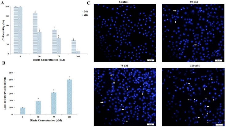
Figure 2.
Effects of rhein on HepaRG cell viability. HepaRG cells were treated with rhein in a series of concentrations (0, 50, 75, 100 μM) for 24 h, 48 h, and 72 h. (A) Cell viability was assessed by the MTT assay. (B) Cell cytotoxicity was measured by the LDH assay. (C) The morphological changes in 24-h rhein-treated HepaRG cells following staining with fluorescent 4′,6-diamidino-2-phenylindole (DAPI) were observed by fluorescence microscopy (Original magnification = 200×, Bar = 50 µm). Arrows indicate bright blue apoptotic cells. Results are the mean ± S.D. (n = 3). LSD t-test was carried out. * p < 0.05, significantly different compared with vehicle control.

