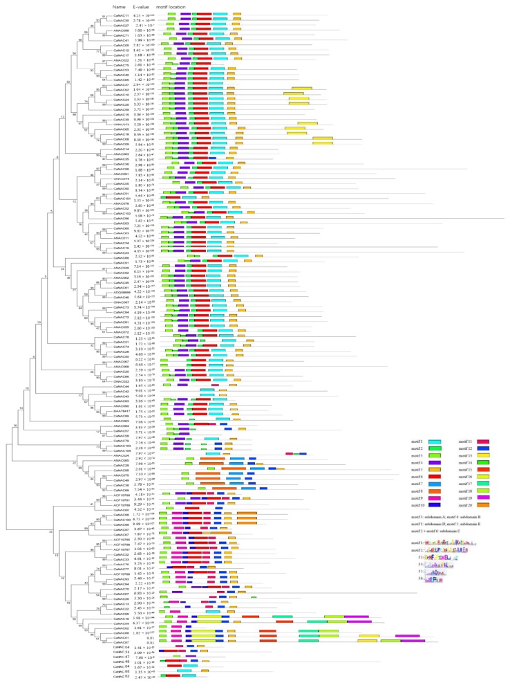
Figure 4.
Alignment of multiple CaNAC and selected ONAC, ANAC domain amino acid sequences, schematic diagram of amino acid motifs of CaNAC and ONAC, ANAC protein groups or subgroups. Motif analysis was performed using Meme 4.12.0 online software (Available online: http://meme-suite.org/tools/meme) as described in the methods. The NAC proteins are listed on the left. The different-colored boxes represent different motifs and their position in each NAC sequence. The sequences of key motifs (motif 1, motif 2, motif 3, motif 4, motif 5, and motif 6) are shown on the bottom right of the figure. A detailed motif introduction for all CaNAC protein is shown in File S2.

