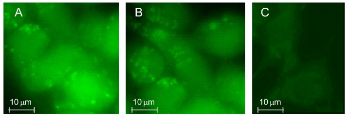
Figure 12.
Rat C6 glioma cells were treated at 37 °C in a 5% CO2 atmosphere with 5 μM of TSPO–FITC–Dex NGs 10 (A) or TSPO-FITC-Dex NGs 11 (B) and the internalized fluorescence was imaged after 24 h of treatment. Images were obtained from 3 to 5 independent experiments and identical fields are presented. Untreated cells and cells treated with 5 μM FITC–Dex alone (after 24 h) were used as a control (C).

