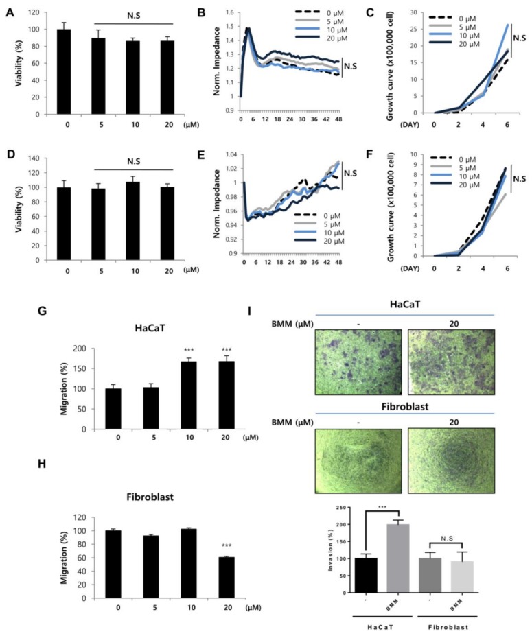
Figure 2.
BMM increases basal layer human keratinocytes (HaCaTs) migration but not proliferation and has no effect on proliferation and migration of dermal fibroblasts (Fbs). (A) Viability of HaCaTs and (D) Fbs. Cells (2 × 10⁴ cell/mL) were seeded and treated with BMM (0–20 μM). After incubation for 24 and 48 h, viability of cells was measured using the MTT solution (5 mg/mL). N.S, not significant; (B) Detection of real time proliferation of HaCaTs and (E) Fbs treated with BMM. Cells (2 × 10⁴ cell/mL) were seeded into eight-well plates pre-coated with 10 mM l-cysteine (Electrode-stabilizing solution). Cells were treated with 5–20 μM of BMM and measurement was started at 48 h by using the ECIS machine. Resistance was calculated with a ratio compared to 0 h. N.S, not significant; (C) Cell counting assay of HaCaT and (F) Fbs treated with BMM (0–20 μM). Cells were seeded (5 × 10³ cells/well) of on six-well plates and the following day, cells were treated with various concentrations of BMM. After two, four, and six days, cells were collected using 1 × Trypsin and counted using a hemocytometer after centrifugation. N.S, not significant; (G) Scratch wound healing assay with HaCaT and (H) Fbs treated as described in (A) 48 h post-treatment. Migration distance was measured using the ImageJ program; (I) Transwell invasion assay with HaCaTs and Fbs treated with 20 μM BMM for 48 h; Invasive ability was measured as described in the Materials and Methods and is quantified under the graph. *** p < 0.001 as compared to that of the control, N.S, not significant.

