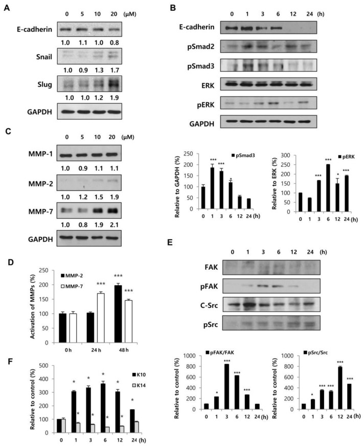
Figure 3.
BMM promotes HaCaT cell migration by inducing an EMT-like phenotype and FAK/Src pathway and stimulates differentiation. (A) HaCaT cells (1 × 10⁵ each) were seeded on 100 mm cell culture dishes and incubated for 24 h; Protein lysates from cells treated with BMM in a dose-dependent and (B) time-dependent manner were collected using RIPA solution, and proteins were resolved by 10% sodium dodecyl sulfate-polyacrylamide gel electrophoresis (SDS-PAGE). Western blot to detect the expression of EMT-related factors was performed, and expression levels were normalized to GAPDH. The values indicate intensities of protein expression with respect to that of the loading control; (C) Western blotting for HaCaT cells treated with BMM for 24 h and MMP expression was measured. The expression levels were normalized to GAPDH. The values indicate intensities of protein expression with respect to that of the loading control; (D) Western blotting for secretion of MMPs from BMM-treated-HaCaT cells. Conditioned medium was concentrated using an Amicon centrifugal filter and total protein concentration was measured by the Bradford assay. The graph indicates expression levels normalized to PonceauS, as a loading control; (E) Western blot to measure the expression of phospho-FAK and phospho-Src as a mechanism of migration in BMM-treated HaCaT cells. The activation of the FAK/Src pathway was calculated by comparing with total-FAK and total-Src; (F) Western blot assay to detect differentiation and proliferation of HaCaT cells, which are respectively marked with K10 and K14, after BMM treatment, and in all cases, expression levels were normalized to GAPDH. * p < 0.05 and *** p < 0.001 as compared to the control.

