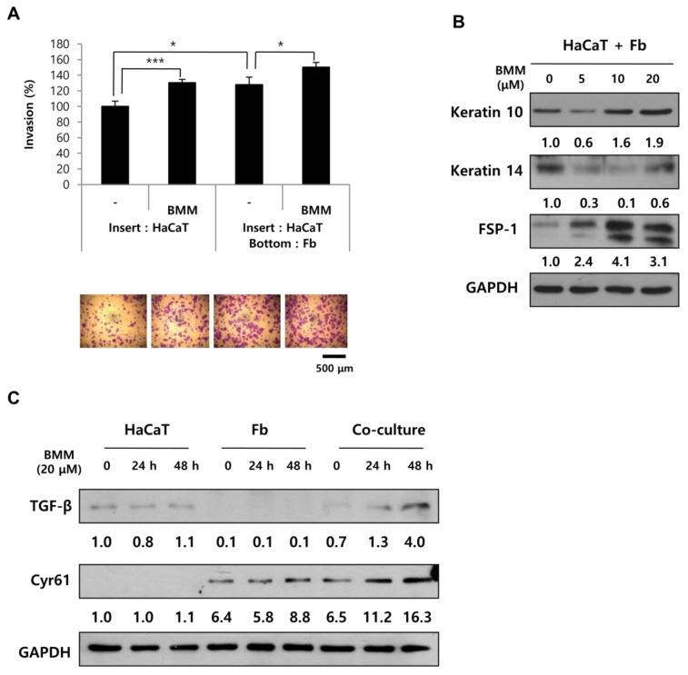
Figure 5.
In co-culture, BMM further stimulates the migratory ability of HaCaT cells through the secretion of TGF-β and Cyr61 from HaCaT and Fbs, respectively. (A) Transwell invasion assay of HaCaT cells after BMM treatment. For single culture, only 7 × 10⁴ cells of HaCaT were seeded on the insert, and an equal number of Fbs were cultured on the bottom at the same time for co-culture. After crystal violet staining, invasive ability was measured using the ImageJ program; (B) Western blotting for co-cultured cells to detect differentiation of HaCaT (K10) or Fbs cells (FSP-1). The values indicate intensities of protein expression with respect to that of the loading control; (C) The expression of TGF-β and Cyr61 on HaCaT, Fbs, and co-cultured cells using western blotting. The values indicate intensities of protein expression with respect to that of the loading control. * p < 0.05 and *** p < 0.001 as compared to the control.

