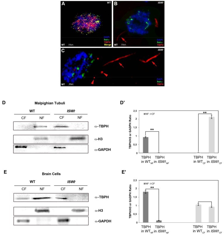
Figure 4.
Loss of ISWI causes severe changes in TBPH localization. (A–C) Compared to WT (A), loss of ISWI dramatically changes TBPH protein (red) distribution between the nucleus and cytoplasm in MT of ISWI- null mutants (B), and higher magnification of other cells in (C). TBPH loses its co-localization with hsrω (green) and it is predominantly distributed in the cytoplasm (red arrows). Moreover, TBPH in the cytoplasm seems to be organized in different trail-like structure (arrows) or dot (arrowheads) (C), (D,E) Cellular fractionation experiments analyzed by Western blots confirming the data for MT cells (D), as well as BCs (E). Anti-GAPDH and anti-H3 were used as internal control to normalize CF and NF. (D’,E’) The intensity of Western blot signals was quantified using ImageJ. The experiment was performed considering five biological replicates. The error bars show the standard deviation. Unpaired Student’s t-test was performed to assay statistical significance; * 0.01 ≤ p-value ≤ 0.05; ** p-value < 0.01.

