Figure 5.
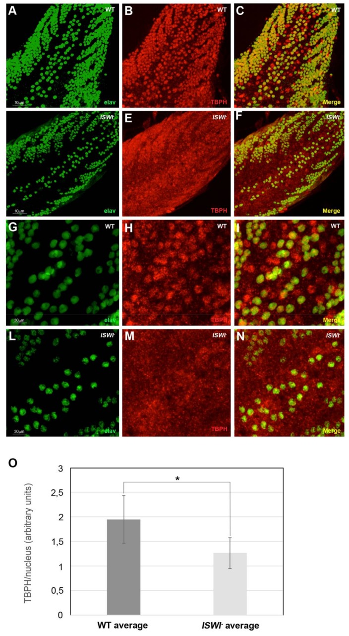
Changes in the distribution of TBPH hnRNP in ISWI null mutant compared to WT in the ventral ganglion of 3rd instar larvae. (A–L) Immunostaining on the ventral ganglia dorsal medial clusters of motoneuron nuclei in third instar wild-type (A–C,G–I magnification) and ISWI- null mutant larvae (D–F,L–N magnification). The mean intensity of TBPH (in red) in the motoneuron nuclei stained with neuronal nuclear protein anti-Elav (green) is significantly reduced in ISWI- null mutants (F,N magnification) compared to wild-type (C,I magnification), as visible also in the merge (C,F). (O) Quantification of the intensity of the immunofluorescence signals. The error bars show the standard deviation. Unpaired Student’s t-test was performed to assay statistical significance; * 0.01 ≤ p-value ≤ 0.05; ** p-value < 0.01.

