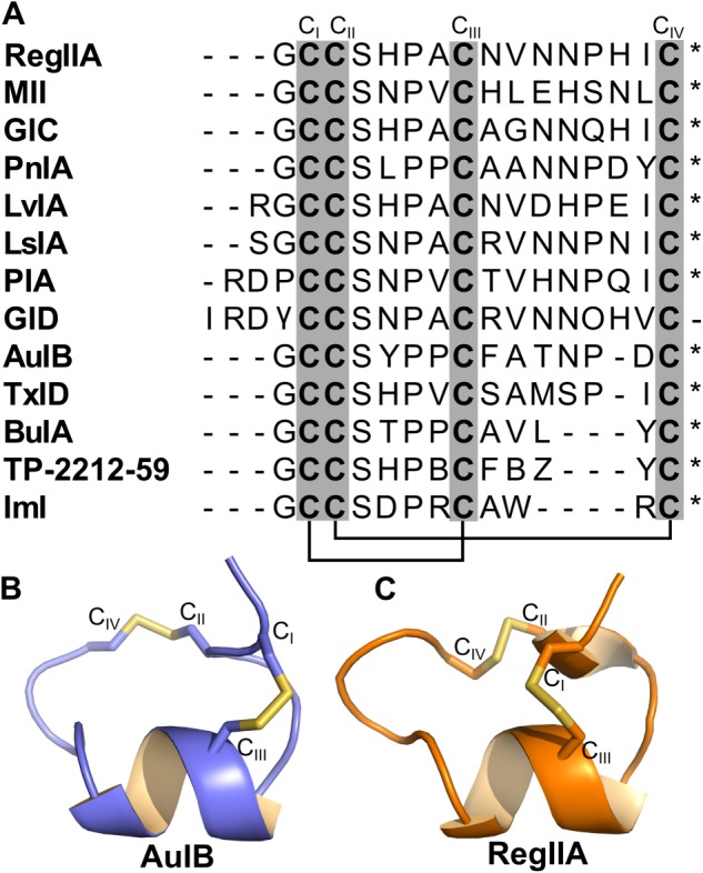
Figure 2.

Sequence and structure of α3β2 and α3β4 nAChR‐targeting α‐conotoxins. (A) Sequence alignment of 13 α‐conotoxins that antagonize α3β2 and α3β4 nAChRs. Cysteine residues CI–CIV (grey columns) form the disulfide bridges between CI–CIII and CII–CIV (black lines) in native α‐conotoxins. * indicates C‐terminal amidation. γ in GID sequence refers to γ‐carboxyglutamate residue. B and Z in the TP‐2212‐59 sequence refer to 2‐aminobutyric acid and norvaline, respectively. (B and C) Structures of α‐conotoxins AuIB and RegIIA respectively.
