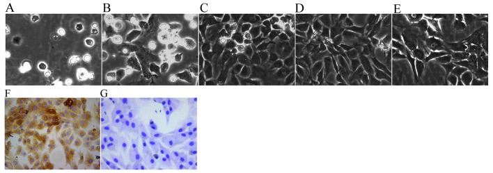
Figure 1.
Morphology and identification of chondrocytes (original magnification, ×200). Primary chondrocytes cultured for (A) 24 h, and (B) 3 and (C) 6 days.(D) First passage chondrocytes cultured for 3 days. (E) Second passage chondrocytes cultured for 3 days. (F) Second passage chondrocytes cultured for 4 days were identified by collagen II immunohistochemistry. (G) Negative control cells also underwent collagen II immunohistochemistry.

