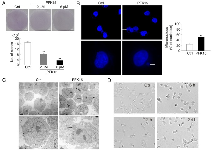
Figure 2.
PFK15 induces an abnormal nuclear morphology. (A) Colony growth assays were performed in RD cells treated with PFK15 (2 and 6 μM). Representative microphotographs of RD cells in adherent conditions after 24 h of treatment in the absence or presence of PFK15 are shown (bar=100 μm). (B) RD cells were cultured for 12 h with PFK15 (6 μM), and immunofluorescence analysis using DAPI was performed to stain nuclei (top panels, magnification ×400; bottom panels, magnification ×1,000). (C) Transmission electron microscopy images of nuclear structures in RD cells are displayed (arrows indicate the micronuclei). (D) RD cells were treated with PFK15 (6 μM) for 24 h, and the morphology of cells were observed by bright field microscopy (magnification, ×40). *P<0.05 vs. control and **P<0.01 vs. control. RD, rhabdomyosarcoma; Ctrl, control.

