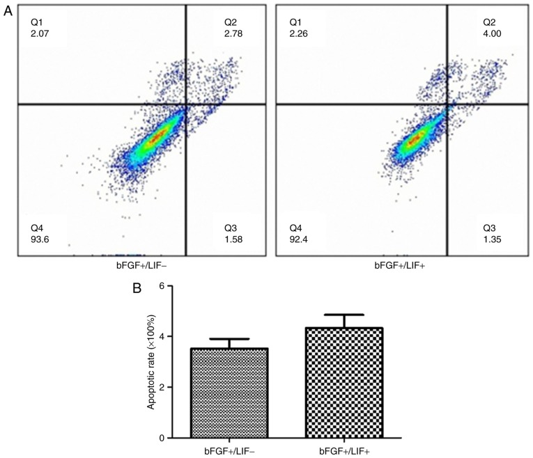
Figure 2.
Effects of LIF on the apoptosis of marmoset iPSCs in culture. (A) Cell apoptosis was analyzed using flow cytometric analysis of FITC Annexin V staining. FITC Annexin V and PI-positive cells were analyzed by flow cytometry. (B) The majority of marmoset iPSCs were FITC Annexin V and PI negative. Apoptosis in the treatment group was 4.81±1.51% and in the control group was 4.25±1.01%. No significant difference in apoptosis was found. Q1, necrotic cells; Q2, late apoptosis; Q3, early apoptosis; Q4, viable cells; LIF, leukemia inhibitory factor; iPSCs, induced pluripotent stem cells; bFGF, basic fibroblast growth factor.

