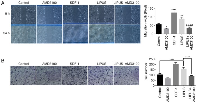
Figure 3.
LIPUS treatment promotes PDLSCs migration. (A) Representative images and quantification from three separate experiments of wound healing assays. (B) Representative images and quantification of transwell migration assays. PDLSCs that penetrated to the lower surface of the membrane were fixed, stained with 0.1% crystal violet, and counted per group. PDLSCs penetrating the membrane were fixed and stained with 0.1% crystal violet after 24 h. Quantification of PDLSCs invasion determined by cell counting. *P<0.05, **P<0.01 and ****P<0.001 vs. control group; ####P<0.001 vs. LIPUS group. LIPUS, low-intensity pulsed ultrasound; PDLSCs, periodontal ligament stem cells; SDF-1, stromal cell-derived factor-1.

