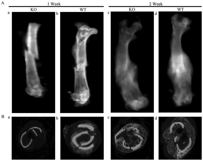
Figure 1.
(A) X-ray images of fractures in (a) PTHKO and (b) PTHWT mice 1 week after fracture. Although fracture lines were clearly visible and a small region of calcification was observed in the surrounding area of fractures in both groups, the calcified region in PTHKO mice was much smaller than that in WT mice. (c) Fracture line remained clear and exogenous callus was relatively small in PTHKO mice 2 weeks after fracture. (d) Blurry fracture line was observed, and the exogenous callus area was clear and relatively large in PTHWT mice 2 weeks after fracture. (B) Coronal micro-computerized tomography views of fractures in (a) PTHKO and (b) PTHWT mice at 1 week and in (c) PTHKO and (d) PTHWT mice at 2 weeks after fracture. PTH, parathyroid hormone; PTHKO, PTH knockout; PTHWT, PTH wild-type.

