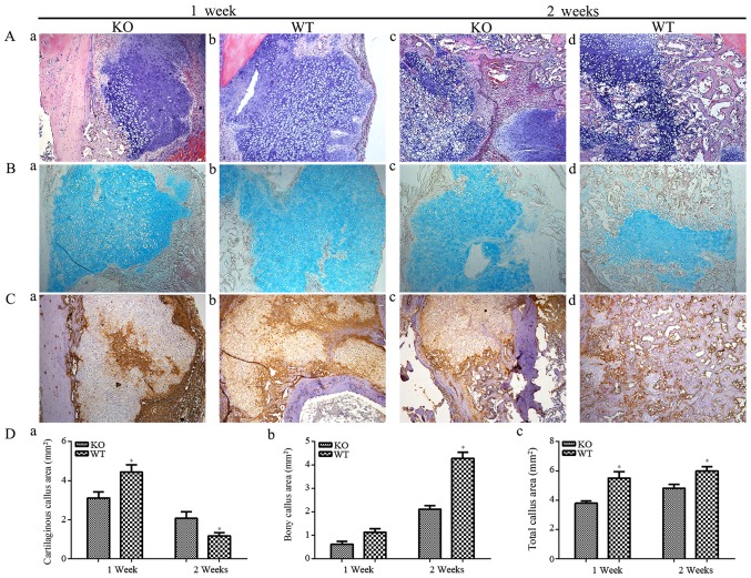
Figure 2.
(A) H&E staining (magnification, ×100) of fractures. (a) In PTHKO mice, fractures developed a smaller cartilaginous callus compared with in (b) WT mice 1 week after fracture. (c) In PTHKO mice at 2 weeks after fracture, little endochondral bone formation and a large cartilage area were detected. (d) In WT mice at 2 weeks after fracture, normal endochondral bone formation and decreased cartilage area were detected. (B) Alcian blue staining (magnification, ×100) of fractures. Light blue area shows cartilaginous callus, which was consistent with the results of H&E staining. (C) Immunohistochemical staining of COL II in fractures (magnification, ×100) in (a) PTHKO and (b) WT mice 1 week after fracture. WT mice exhibited strong positive staining for COL II; however, COL II expression was detected only in the central region of the cartilage expansion area in PTHKO mice. Immunohistochemical staining of COL II in (c) PTHKO and (d) WT mice 2 weeks after fracture. COL II expression was slightly increased in PTHKO mice, but remained lower than that in WT mice. (D) Statistical analysis of (a) cartilaginous callus area, (b) bony callus area and (c) total callus area. *P<0.05. PTH, parathroid hormone; PTHKO, PTH knockout; WT, wild-type.

