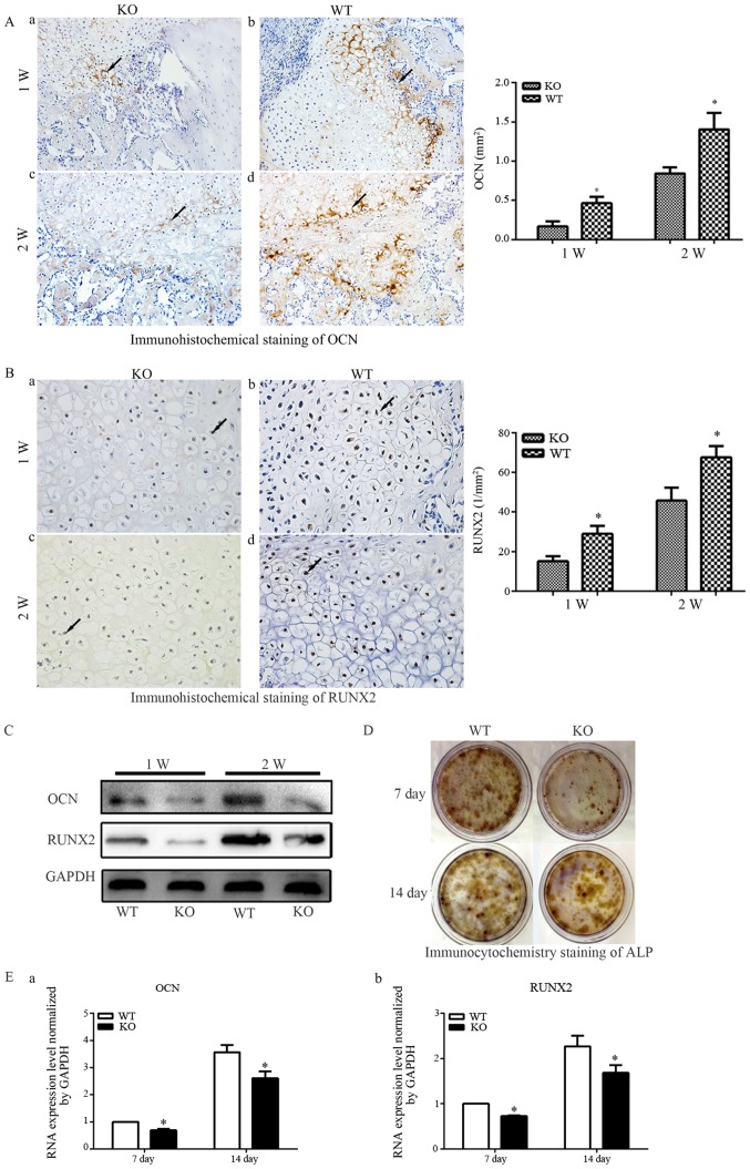
Figure 3.
(A) Immunohistochemical staining of OCN (magnification, ×200) in fractures of (a) PTHKO and (b) WT mice 1 week after fracture. OCN expression was significantly lower in PTHKO mice compared with in WT mice and was primarily distributed around the edge of the callus. Immunohistochemical staining of OCN in fractures of (c) PTHKO and (d) WT mice 2 weeks after fracture. OCN expression was significantly reduced in PTHKO mice compared with in WT mice; however, it was increased in both groups compared with after 1 week. (B) Immunohistochemical staining of RUNX2 (magnification, ×400) in fractures of (a) PTHKO and (b) WT mice 1 week after fracture. RUNX2 expression was significantly lower in PTHKO mice compared with in WT mice, although it was relatively strong in the area surrounding the cartilaginous callus in both groups. Immunohistochemical staining of RUNX2 in fractures of (c) PTHKO and (d) WT mice 2 weeks after fracture. RUNX2 was strongly expressed in WT mice, whereas RUNX2 expression remained lower in PTHKO mice compared with in WT mice, and was mainly concentrated in the outer periphery of the cartilaginous callus. Black arrows indicate positive areas. (C) Callus proteins, including OCN and RUNX2, were detected using western blot analysis. (D) ALP immunocytochemistry. Reduced ALP staining intensity was detected in PTHKO mice compared with in WT mice. (E) Reverse transcription-quantiative polymrease chain reaction analysis of mRNA expression of (a) OCN and (b) RUNX2 in BMSC-derived osteoblasts; the mRNA expression levels were significantly lower in PTHKO mice compared with in WT mice 1 and 2 weeks after induction. *P<0.05. ALP, alkaline phosphatase; BMSC, bone marrow mesenchymal stem cells; OCN, osteocalcin; PTH, parathroid hormone; PTHKO, PTH knockout; RUNX2, runt-related transcription factor 2; WT, wild-type.

