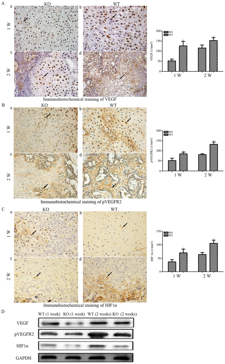
Figure 5.
(A) Immunohistochemical staining of VEGF-A (magnification, ×400) in fractures. (a) In PTHKO and (b) WT mice 1 week after fracture, VEGF-A expression was significantly reduced in PTHKO mice compared with in WT mice. (c) In PTHKO and (d) WT mice 2 weeks after fracture, VEGF-A expression was increased in PTHKO mice, but remained significantly lower than that in WT mice. (B) Immunohistochemical staining of pVEGFR2 (magnification, ×400) in fractures. (a) In PTHKO and (b) WT mice 1 week after fracture, a significantly smaller number of pVEGFR2-positive cells was detected in the cartilaginous callus in PTHKO mice compared with in WT mice. (c) In PTHKO and (d) WT mice 2 weeks after fracture, a large number of pVEGFR2-positive cells was observed in the cartilaginous callus in WT mice, whereas a much lower level of angiogenesis was detected in PTHKO mice. (C) Immunohistochemical staining for HIF1α (magnification, ×400) in fractures. (a) In PTHKO and (b) WT mice 1 week after fracture, the expression levels of cytoplasmic HIF1α were significantly lower in PTHKO mice. (c) In PTHKO and (d) WT mice 2 weeks after fracture, HIF1α expression was increased in both groups; however, the expression remained lower in PTHKO mice compared with in WT mice. Black arrows indicate positive areas. (D) Protein expression levels of VEGF, pVEGFR2 and HIF1α were detected by western blot analysis. HIF1α, hypoxia inducible factor-1α; PTH, parathroid hormone; PTHKO, PTH knockout; pVEGFR, phosphorylated-VEGF receptor 2; VEGF, vascular endothelial growth factor; WT, wild-type.

