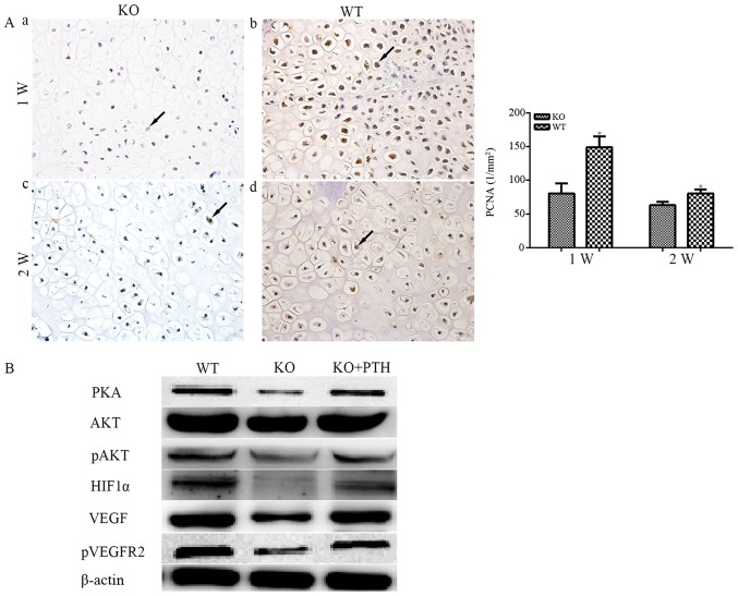
Figure 6.
(A) Immunohistochemical staining of PCNA (magnification, ×400) in fractures. (a) In PTHKO and (b) WT mice 1 week after fracture, rapid cell proliferation was observed, whereas PTHKO mice exhibited a significant reduction in PCNA-positive rate compared with in WT mice. (c) In PTHKO and (d) WT mice 2 weeks after fracture, cell proliferation in WT mice remained faster than that in PTHKO mice; however, it slowed down in both groups. Black arrows indicate positive areas. *P<0.05. (B) Western blot analysis detected reduced expression of PKA/pAKT/HIF1α/VEGF, and a lower level of pVEGFR2 in PTHKO BMSC-derived osteoblasts 2 weeks after induction. Addition of exogenous PTH in the culture medium partially reversed downregulation of the PKA/pAKT/HIF1α/VEGF pathway. AKT, serine/threonine protein kinase; BMSC, bone marrow mesenchymal stem cell; HIF1α, hypoxia inducible factor-1α; pAKT, phosphorylated-AKT; PCNA, proliferating cell nuclear antigen; PKA, protein kinase A; PTH, parathroid hormone; PTHKO, PTH knockout; pVEGFR2, p-VEGFR2; VEGF, vascular endothelial growth factor; VEGFR2, VEGF receptor 2; WT, wild-type.

