Figure 6.
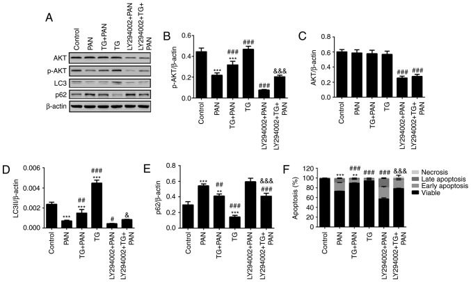
TG promotes the activation of autophagy by PI3K signaling during podocyte injury. (A) Representative bands of AKT, p-AKT, LC3 and p62 proteins in the individual groups. Quantitative evaluation of the levels of (B) p-AKT, (C) AKT, (D) LC3 and (E) p62 in the individual groups. The expression of p-AKT was markedly inhibited by LY294002. The expression of p-AKT was markedly increased in the TG+PAN and LY294002+TG+PAN groups, compared with the expression in the PAN group and LY294002+PAN group, respectively. TG promoted the expression of LC3II and decreased the expression of p62. (F) Apoptotic rates in the groups. Data are presented as the mean ± standard deviation (n=3). **P<0.01 and ***P<0.001 vs. control group; #P<0.05, ##P<0.01 and ###P<0.001 vs. PAN group; &P<0.05 and &&&P<0.001 vs. LY294002+PAN group. TG, tripterygium glycoside; PAN, puromycin aminonucleoside; PI3K, phosphatidylinositol 3-kinase; p-, phosphorylated; LC3, microtubule-associated protein 1A/1B-light chain 3.

