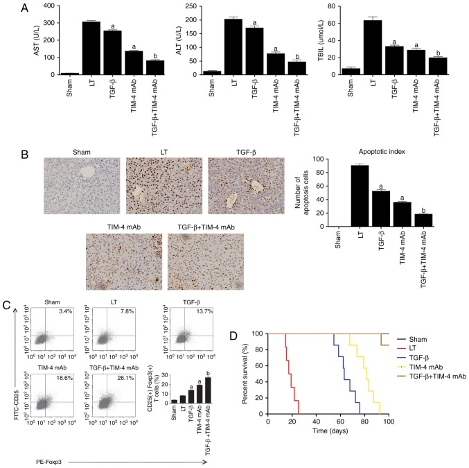
Figure 5.
The effect of TIM-4 blockade with exogenous TGF-β injection on acute reaction response in vivo. (A) Mice were injected with either anti-TIM-4 mAb (0.35 mg/mouse), 0.5 ml TGF-β (1 ng/ml) or in combination via murine portal veins to establish LT (LT group were treated with PBS as control). Treatment in each group was administered continuously at the same dose for a total of 2 days following surgery. Levels of AST, ALT and TBIL were determined in a clinical biochemical laboratory on day 7 following surgery. (B) Hepatocyte apoptosis was detected using the terminal deoxynucleotidyl transferase-mediated 2′-deoxyuridine 5′-triphosphate nick-end labeling method (magnification ×400). (C) T cells were purified from hepatic tissue. Following washing and re-suspension, cells were stained with PE-Foxp3 and FITC-CD25 antibodies and acquired for fluorescence-activated cell sorting analysis. (D) Mice survival time was observed and analyzed using the Log-rank test. Data are presented as the mean ± standard deviation of the mean. aP<0.05 vs. LT group; bP<0.05 vs. TGF-β and TIM-4 mAb groups. TIM-4, T cell immunoglobulin-domain mucin-domain-4; TGF-β, transforming growth factor-β; mAb, monoclonal antibodies; LT, liver transplantation; AST, aspartate aminotransferase; ALT, alanine aminotransferase; TBIL, total bilirubin in serum; Foxp3, forkhead box P3; FITC, fluorescein isothiocyanate; CD, cluster of differentiation; PE, phycoerythrin.

