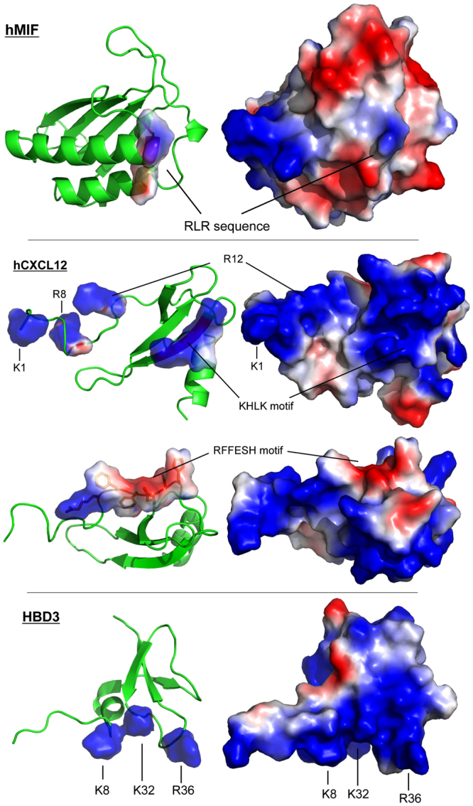
Figure 8.

Comparison of the surface charges of MIF, CXCL12, and human β-defensin-3. Comparison of the three-dimensional structures of MIF, CXCL12, and human β-defensin-3 (HBD3) focusing on the surface charges. The relevant positively charged residues, the RLR, and the KHLK sequence, as well as the RFFESH motif are indicated as shown. Blue, positively charged residues arginine, lysine, or histidine; red, negatively charged residues aspartate and glutamate. Protein structures were produced/visualized with PyMOL.
