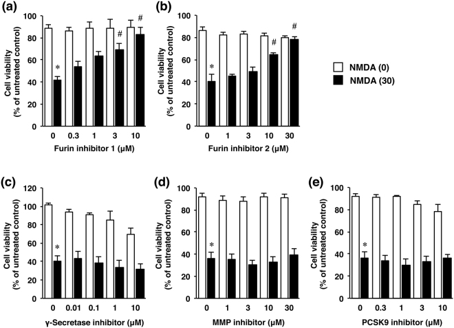
Figure 3.
Effects of furin inhibitors (a,b) as well as several protease inhibitors (c–e) on NMDA-induced cell injury. Cell viability in cultures of cortical neurons treated with 0 µM (white bars) or 30 µM NMDA (black bars) without or with the indicated concentrations of furin inhibitor 1 (a), furin inhibitor 2 (b), γ-secretase inhibitor (c), MMP inhibitor (d) or PCSK9 inhibitor (e). The relative cell viability was expressed as the percentage of the absorbance at 450 nm of each treatment group against that of the untreated control group. Results are the means ± SE (n = 4 independent experiments). *Indicates a significant difference from the NMDA- and corresponding inhibitor-untreated group (p < 0.05); and #, a significant difference from the NMDA-treated and furin inhibitor 1- untreated group (p < 0.05).

