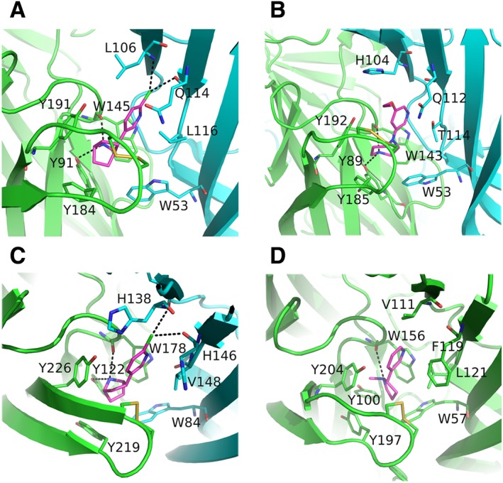
Figure 2.

Close views of wild‐type or chimeric nAChR ligand‐binding sites. (A) The α7‐AChBP bound to epibatidine (PDB ID: 3SQ9) (Li et al., 2011). (B) Engineered AChBP towards α4/α4 bound to NS3920 (PDB ID: 4UM3) (Shahsavar et al., 2015). (C) The α2‐ECD bound to epibatidine (PDB ID: 5FJV) (Kouvatsos et al., 2016). (D) The α4β2 nAChR bound to nicotine (PDB ID: 5KXI) (Morales‐Perez et al., 2016). The principal sides are shown in green, the complementary in cyan and the agonists in magenta. Interactions are shown in black dashed lines. The coordinates of all the structures depicted were retrieved from Protein Data Bank (http://www.wwpdb.org), and PyMol (http://www.pymol.org) was used to generate the figures.
