
Figure 2.
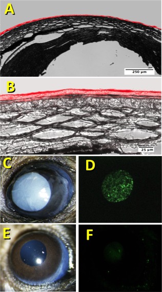
Adherence of compressed collagen gel to mouse cornea. (A): Fluorescently labeled CCG was adhered to the surface of a mouse cornea ex vivo as described under Methods. Cryosections of the cornea were imaged using bright‐field + fluorescent illumination to illustrate the close adherence of the CCG to the corneal surface. (B): Corneal section in (A) with a higher magnification. (C): A 2 mm diameter CCG with embedded DiO‐labeled CSSC was attached to a mouse cornea in vivo after corneal wounding as described in “Materials and Methods” section. (D): Fluorescent image of the eye in (C) revealing the fluorescent CSSC in the gel. (E): Bright field image of the mouse eye from (C) 48 hours after attachment of the CCG. (F): Fluorescent image of the eye in (E) illustrating lack of CSSC associated with the cornea. Abbreviations: CCG, compressed collagen gel; CSSC, corneal stromal stem cells.