Figure 3.

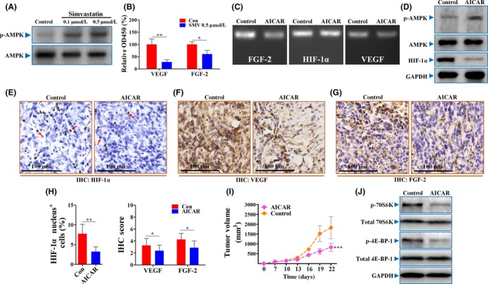
Simvastatin (SMV) reduced hypoxia‐inducible factor‐1α (HIF‐1α) protein levels by activation of AMP kinase (AMPK). A, Representative Western blot analysis showing AMPK/phosphorylated (p‐)AMPK protein levels in 4T1 tumor cells treated with PBS or different doses of SMV. B, Quantifications of secreted vascular endothelial growth factor (VEGF) and fibroblast growth factor‐2 (FGF‐2) proteins in supernatants of 4T1 cells by ELISA (n = 6). C, Representative images showing the mRNA levels of HIF‐1α, VEGF, and FGF‐2 in 4T1 cells untreated or treated with 1 mmol/L 5‐aminoimidazole‐4‐carboxamide ribonucleotide (AICAR). D, Immunoblotting for p‐AMPK/AMPK/HIF‐1α in AICAR‐treated or ‐untreated 4T1 cells. GAPDH was used as a control for protein loading. Immunohistochemical (IHC) detection of HIF‐1α (E), VEGF (F), and FGF‐2 (G) in sections of 4T1 tumors. Red arrows in panel (E) indicate HIF‐1α nucleus+ cells. Scar bar = 100 μm. H, Percentage of HIF‐1α nucleus+ cells (% total cells; left) and IHC quantification of VEGF and FGF‐2 in sections of 4T1 tumors treated with PBS (Con) or 400 mg/kg AICAR (right; n = 8). I, Representative images showing growth curve of 4T1 tumors from mice untreated or treated with AICAR (n = 8). J, Immunoblotting for both total and phosphorylation levels of 70S6K and 4E‐BP‐1, two direct targets of mTOR, in AICAR‐treated or ‐untreated 4T1 cells. Quantitative data are indicated as mean ± SD. *P < .05; **P < .01; ***P < .001. ns, no statistically significant difference (P > .05)
