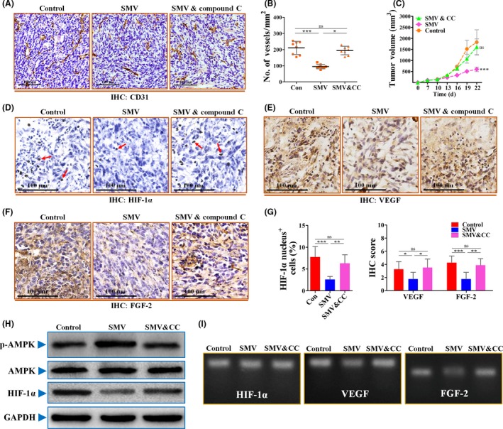
Figure 6.

Inhibition of AMP kinase (AMPK) activation by compound C abrogated the inhibitory effects of simvastatin (SMV) on hypoxia‐inducible factor‐1α (HIF‐1α)‐induced tumor angiogenesis. A, Representative images for showing immunohistochemical (IHC) analysis of CD31+ vessels in 4T1 tumors from mice treated with PBS (Control; Con), 15 mg/kg SMV, or combined treatment with 15 mg/kg SMV and 20 mg/kg compound C (CC). Scale bar = 100 μm. B, Quantification of microvessel density (no. of vessels/mm2) in 4T1 tumors (n = 8). C, Representative images showing growth curve of 4T1 tumor from mice untreated or treated with SMV or combination treatment of SMV and CC (n = 8). D‐F, Representative images of immunostaining for HIF‐1α (D), vascular endothelial growth factor (VEGF) (E), and fibroblast growth factor‐2 (FGF‐2) (F) in 4T1 tumor sections of control, SMV, or SMV and CC combination groups. Red arrows in panel (D) indicate HIF‐1α nucleus+ cells. Scar bar = 100 μm. G, Percentage of HIF‐1α nucleus+ cells (% total cells; left) and quantification of IHC scores of VEGF and FGF‐2 in sections of 4T1 tumors (right; n = 8). H, Immunoblotting for phosphorylated p‐AMPK, total AMPK, and HIF‐1α of 4T1 cells treated with PBS (Control), 0.5 μmol/L SMV, or combination of 0.5 μmol/L SMV and 5 μmol/L CC. GAPDH was used as a control for protein loading. I, RT‐PCR for determining the mRNA levels of HIF‐1α, VEGF, and FGF‐2 of 4T1 tumor cells. Quantitative data are indicated as mean ± SD. *P < .05; **P < .01; ***P < .001. ns, no statistically significant difference (P > .05)
