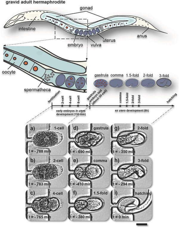
Figure 3.

Study of complete C. elegans embryogenesis on‐chip. Schematic of anatomical structures of a gravid C. elegans hermaphrodite with two bilaterally symmetric gonad arms that are connected to a central uterus through the spermatheca. Embryos are shaded in blue‐gray. The embryonic development timeline shows several key events with distinguishable morphological changes during in utero (one‐cell to gastrula, ≈30 cells) and ex utero development (gastrula to hatching). a–i) Live imaging of normal on‐chip embryogenesis from the first division to larva hatching (T = 23 °C). Bright‐field images (50×/0.55 objective) of a microfluidic trap located in between two parallel sections of the serpentine channel in the trapping array (see Figure 2). a) An immobilized C. elegans embryo is shown in the one‐cell stage (a), and b–i) in subsequent main embryonic development phases. Note that this particular embryo was trapped with the anterior pole on the left. The moment of hatching defines t = 0 min. This sequence was taken over a timespan of ≈13 h (at the time points indicated in the figure), demonstrating reliable embryo positioning over prolonged periods. Scale bar = 20 µm.
