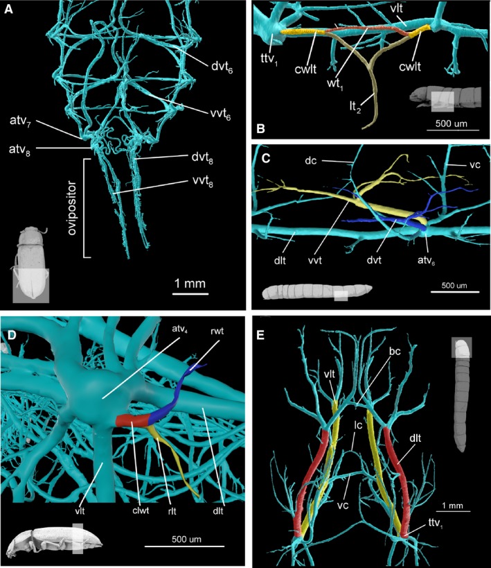
Figure 7.

Anatomy of tracheal system. (A) Abdomen of imago, dorsal view. Specimen has extruded ovipositor and aerating trachea are pulled with it. (B) Mesothorax of larva lateral view. In colour, mesoleg (brown) and fore wing trachea (red) and common wing leg trachea (yellow). (C) Abdomen of larva, segment 5, dorsal view. In colour, dorsal visceral trachea (blue) and ventral visceral trachea (blue). (D) Tracheal vestibule of abdominal segment 4 of imago, lateral view. In colour, leg homologues trachea (yellow), wing homologues trachea (blue) and common wing leg homologue trachea (red). (E) Head and prothorax of larva, dorsal view. In color, longitudinal tracheal trunks, dorsal (red) and ventral (yellow). atv4, tracheal vestibule of 4 abdominal segment; atv6–atv8, tracheal vestibule of 6–8 abdominal segment; bc, brain commissure; lwt, common leg‐wing trachea; cwlt, common leg‐wing trachea; dc, dorsal commissure; dlt, dorsal longitudinal trunk; dvt, dorsal visceral trachea; dvt6 and dvt8 – dorsal visceral trachea of 6 and 8 abdominal segment; lc, proleg commissure; lt1, proleg trachea; rlt, rudimentary leg trachea; rwt, rudimentary wing trachea; ttv1, tracheal vestibule of mesothorax; vc, ventral commissure; vlt, ventral longitudinal trunc; vvt, ventral visceral trachea; vvt6 and vvt8 – ventral visceral trachea of 6 and 8 abdominal segment; wt1, fore wing trachea.
