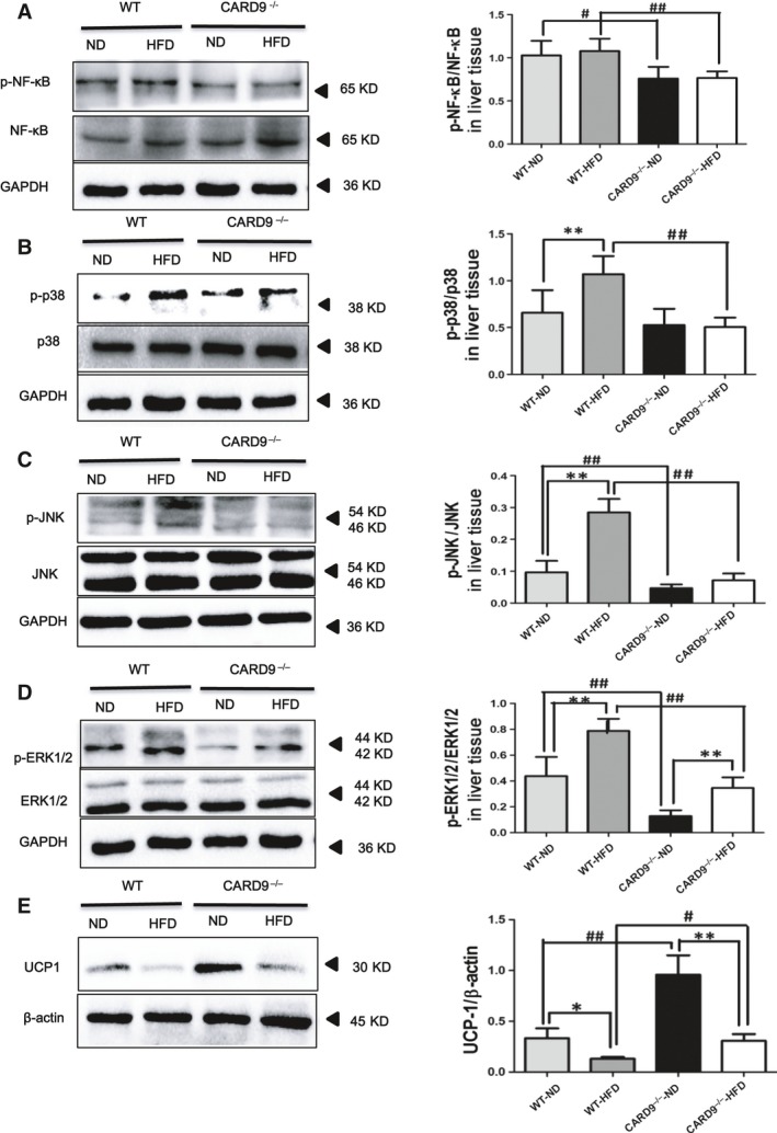
Figure 7.

Protein expression of inflammation and MAPK signalling pathways in the liver of the CARD9−/− and WT mice after ND and HFD feeding, along with the UCP‐1 expression in BAT. Western blotting for phosphorylated NF‐κB (P‐NF‐κB)/total NF‐κB (A). Western blotting of signalling molecules involved in MAPK pathway [P‐p38/p38, (B); P‐JNK/JNK, (C); P‐ERK/ERK, (D)]. Western blotting of UCP‐1 (E). *P < 0.05, **P < 0.01, HFD vs. ND; # P < 0.05, ## P < 0.01, CARD9−/− vs. WT.
