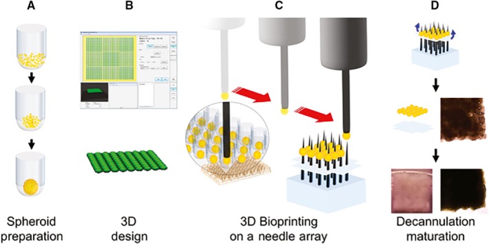
Figure 2.

Schematic of biomaterial‐free bioprinting of a cardiac patch. A, Cells (CM, FB, EC) are aggregated in ultra‐low attachment 96‐well plates to form spheroids. B, The desired 3D structure is designed using computer software. C, The robot picks up individual spheroids using vacuum suction and loads them onto a needle array. D, Spheroids are allowed to fuse. The 3D bioprinted cardiac tissue is then removed from the needle array and further cultured to allow the needle holes to be resorbed (reproduced with permission from Ong et al 56)
