
Figure 2.
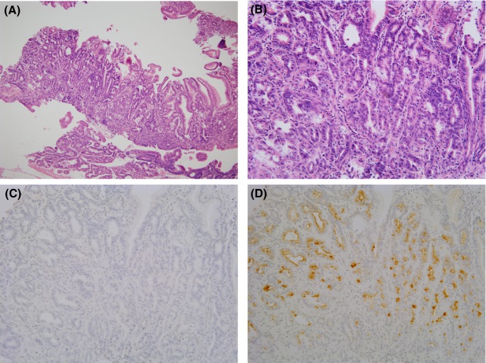
Primary tumor from which cell lines were established. (A) Hematoxylin and eosin staining (10×). (B) Higher magnification (40×). Tumor shows a well‐differentiated phenotype. (C) Pepsinogen 1 was negative in the tumor. (D) Class III mucin was positive in the tumor and thus the tumor was classified as a pyloric phenotype