
Figure 6.
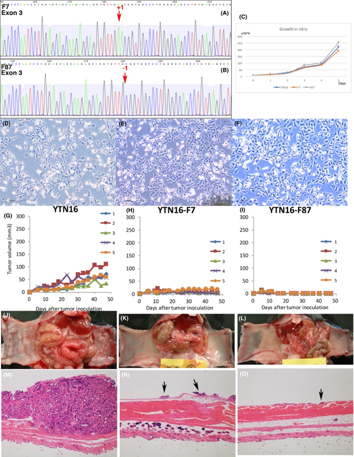
Fgfr4 disrupted YTN16. (A) F7 has an insertion of adenine in exon 3 of Fgfr4, and Fgfr4 is disrupted by a frameshift termination. (B) F87 has a deletion of adenine in exon 3 of Fgfr4, and Fgfr4 is disrupted by a frameshift termination. (C) Growth rates of YTN16, F7 and F87 were not different in vitro. (D‐F) Microscopic morphology of YTN16, F7, and F87 showed no difference in vitro. (G‐I) F7 and F87 do not form s.c. tumors. (J‐L) F7 and F87 formed very small foci of peritoneal dissemination in 2 out of 5 mice. YTN16 formed large nodules in 5 out of 5 mice in the same period. (M‐O) YTN16 formed large nodules of peritoneal dissemination. F7 and F87 formed only very small peritoneally disseminated tumors in macroscopically positive mice only. Arrows: small peritoneally disseminated tumor