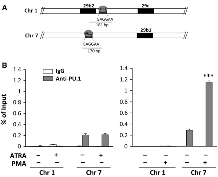
Figure 2.

In vivo interaction of PU.1 with miR‐29b promoters. (A) Schematic representation of the putative PU.1 binding sites within the human miR‐29b2/c promoter on chromosome 1q32.2 and within the human miR‐29a/b1 promoter on chromosome 7q32.3. (B) Analysis of in vivo recruitment of PU.1 to both miR‐29b promoters performed by chromatin immunoprecipitation with an antibody directed against PU.1 in Kasumi‐1 cells treated with all‐trans‐retinoic acid (ATRA) or phorbol 12‐myristate 13‐acetate (PMA). The data are shown as percentage of the Input (genomic DNA collected before immunoprecipitation). IgG: negative control. Values represent the means of 3 separate experiments ±SD. ***P < .001 compared to untreated cells
