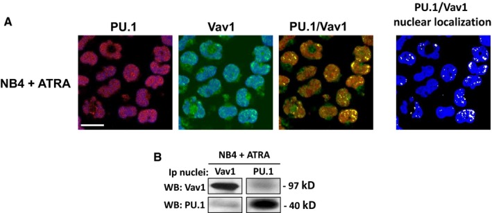
Figure 4.

Vav1 and PU.1 association in all‐trans‐retinoic acid (ATRA)‐treated NB4 cells. (A) Representative confocal images of NB4 cells treated with ATRA and stained with antibodies against Vav1 (green staining) and PU.1 (red staining). TO‐PRO ®‐3 Stain was used to counterstain the nucleus (shown in blue). PU.1 and Vav1 images are shown as the overlay of the protein staining (red or green) with the staining of the nucleus (blue). Merged PU.1/Vav1 staining is shown with colocalization resulting in yellow. To the right, PU.1/Vav1 colocalization points were white coloured and overlapped to nuclear staining (blue). Bar = 20 μm. (B) Representative Western blot analysis with the indicated antibodies of Vav1 and PU.1 immunoprecipitates from nuclei of NB4 cells treated with ATRA. The data are representative of 3 separate experiments
