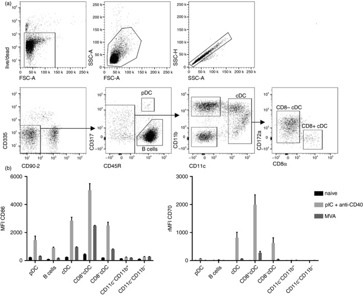
Figure 1.

Up‐regulation of CD86 and CD70 on splenic antigen‐presenting cells (APCs) after modified vaccinia virus Ankara (MVA) immunization. C57BL/6 mice were immunized intravenously (i.v.) with 5 × 10⁷ TCID 50 modified vaccinia virus Ankara–Bavarian Nordic® (MVA‐BN ®) or pIC + anti‐CD40. Expression of CD86 and CD70 on splenic APC subsets was analysed after 41 hr by 11‐colour flow cytometry. Cells were gated as shown in the exemplary dot plots (a) and were defined as follows: plasmacytoid dendritic cells (live CD90.2− CD335− CD45R+ CD317+), B cells (live CD90.2− CD335− CD45R+ CD317−), conventional dendritic cells (live CD90.2− CD335− CD45R− CD317− CD11c+), CD8+ conventional dendritic cells (live CD90.2− CD335− CD45R− CD317− CD11c+ CD172a− CD8α+), CD8− conventional dendritic cells (live CD90.2− CD335− CD45R− CD317− CD11c+ CD172a+ CD8α−), CD11c− CD11b+ ((live CD90.2− CD335− CD45R− CD317− CD11c− CD11b+) and CD11c− CD11b− (live CD90.2− CD335− CD45R− CD317− CD11c− CD11b−). (b) Bar graphs show the mean fluorescence intensity (MFI) ± SEM of CD86 and the relative (r)MFI of CD70 of three mice per group. The rMFI was calculated by subtracting the MFI of the fluorescence‐minus‐one control from the MFI of stained samples. Data are representative of three independent experiments for MVA‐BN ® and one for pIC + anti‐CD40.
