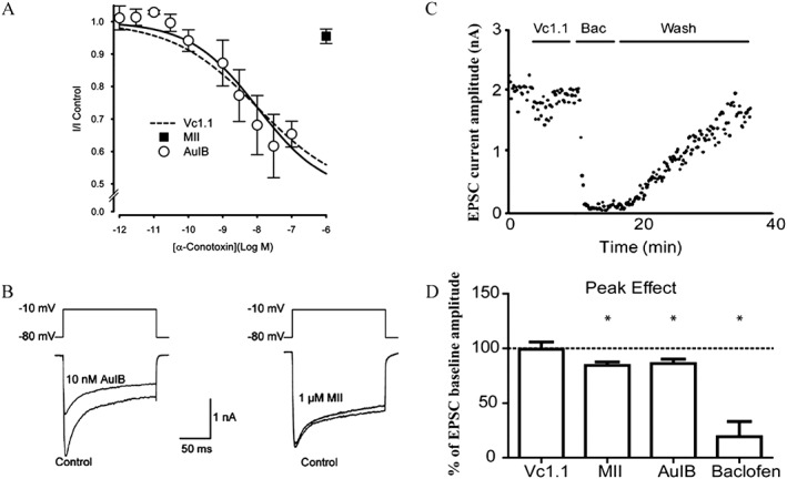
Figure 1.

Effects of analgesic α‐Ctxs on VGCC currents in DRG neurons and EPSCs in spinal cord neurons. An alternative hypothesis is that α‐Ctxs exert their analgesic effects by inhibition of VGCCs via activation of GABAB receptors rather than by inhibition of α9α10 nAChRs. (A and B) Examples of experiments that demonstrate α‐Ctx activity on GABAB receptors, whereas (C) and (D) show experiments demonstrating a lack of α‐Ctx activity on GABAB receptors. (A) Concentration–response relationship for inhibition of VGCC currents by Vc1.1, AuIB and MII in cultured rat DRG neurons (figures used with permission of Klimis et al., 2011). Similar activity has been reported for analogues of Vc1.1 and for RgIA (see text for full discussion and references). One study did not replicate key findings for Vc1.1 or RgIA in DRG neurons (Wright et al., 2015) (data not shown). (B) AuIB, but not MII, inhibits VGCC currents in DRG neurons. These findings support the hypothesis that the analgesic effects of some α‐Ctxs are mediated through activation of GABAB receptors. However, Vc1.1 failed to inhibit EPSCs in spinal cord neurons via activation of presynaptic GABAB receptors on DRG afferents. (C) Electrophysiological recordings from rat spinal cord neurons in a slice preparation showing the effect of Vc1.1 and baclofen (Bac) on EPSC amplitudes. (D) Histogram of EPSCs in the presence of α‐Ctxs or baclofen compared with baseline controls. Note lack of effect of Vc1.1 and small inhibitory effects of AuIB and MII (the latter attributed to block of α3‐containing nAChRs). The GABAB receptor agonist baclofen is shown as a positive control (figures used with permission, Napier et al., 2012).
