Figure 1.

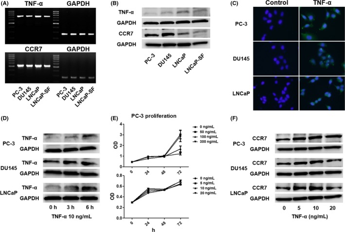
Low‐dose tumor necrosis factor‐α (TNF‐α) induces C‐C chemokine receptor 7 (CCR7) expression in prostate cancer cells. A,B, Total RNA and protein were extracted from prostate cancer cells, and their mRNA and protein levels were analyzed using RT‐PCR A and Western blotting B. C, Prostate cancer cells (1.0 × 10⁵ cells/well) were seeded into 6‐well plates and cultured until they reached 60%‐70% confluence. The cells were incubated with a primary anti‐TNF‐α antibody, followed by incubation with a secondary antibody conjugated with FITC (green). Cells were counterstained with DAPI (blue). D, Changes in TNF‐α protein levels in prostate cancer cells after stimulation with exogenous TNF‐α (10 ng/mL) were determined by Western blotting. E, PC‐3 cell proliferation was determined with the WST‐1 assay using a range of TNF‐α concentrations. Although high TNF‐α concentrations (100 and 300 ng/mL, compared with 0 ng/mL, P < .01) significantly inhibited cell proliferation at 72 hour (upper panel), low concentrations did not lead to inhibition of cell proliferation (lower panel). Data are presented as mean ± SD. F, Prostate cancer cells were treated with TNF‐α at different concentrations for 6 hour, and Western blot analysis was used to detect CCR7
