
Figure 1.
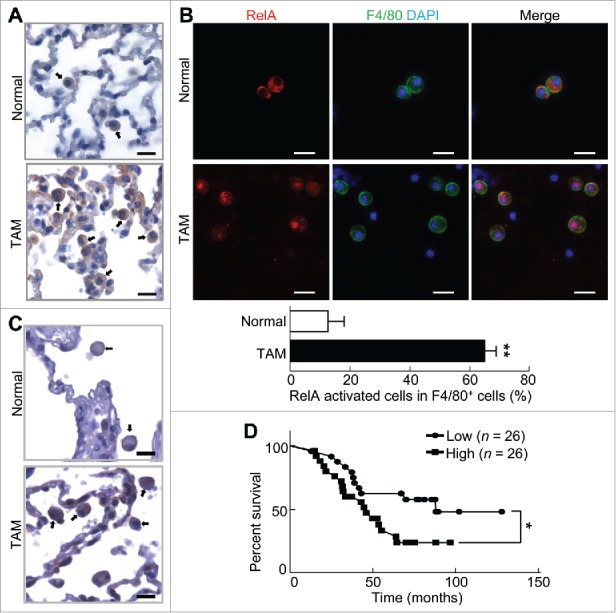
Myeloid RelA activation is associated with poor lung cancer patient survival. (A) IHC staining of RelA in lung tissues from untreated mice or mice with lung tumors induced by urethane. Arrows indicate myeloid cells. Scale bar: 20 μm. (B) IF staining of RelA and F4/80 in BAL cells from untreated mice or mice with lung tumors induced by urethane. Scale bar: 20 μm. Data are means ± SD (n = 3 mice, 3–5 images per mouse). (C) IHC staining of RelA in human lung cancer tissues and matched normal human lung tissues. Arrows indicate myeloid cells. Scale bar: 20 μm. (D) Kaplan-Meier survival curve showing the association between pulmonary myeloid RelA activation and lung cancer patient survival. p = 0.033 (Log-rank test); p = 0.048 (Gehan-Breslow-Wilcoxon test); p = 0.046 (Cox proportional hazards model).