Figure 1.

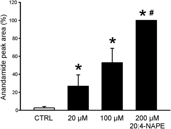
Anandamide concentration after 20:4‐NAPE application to spinal cord slices. Three different concentrations of 20:4‐NAPE (20, 100 and 200 μM) were applied to spinal cord slices. Increasing content of anandamide was detected in the extracellular solution after 20:4‐NAPE application, in a concentration dependent manner. *P < 0.05, significantly different from control, # P < 0.05, significantly different from 20 and 100 μM 20:4‐NAPE; repeated measures ANOVA on ranks followed by Student–Newman–Keuls test; n = 5).
