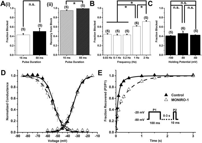
Figure 5.

MONIRO‐1 block of human Cav3.1 channels. (A) (i) Bar graph showing fraction blocked and (ii) recovery from block by 3 μM MONIRO‐1, with a depolarizing pulse duration of 15 ms or 50 ms, from a holding potential of −80 mV repeated every 10 s. The number of cells is indicated in parentheses. (B) Bar graph of the fractional current inhibited by 3 μM MONIRO‐1 at five different frequencies (0.03, 0.1, 0.2, 1 and 2 Hz; holding potential −80 mV). The number of cells is indicated in parentheses. (C) Effect of holding potential (−100, −80 and −60 mV) on the fraction of current inhibited by 3 μM MONIRO‐1 (pulsed at 0.1 Hz). The number of cells is indicated in parentheses. (D) Activation and SSI curves in the absence (control) and presence of 3 μM MONIRO‐1. Activation curves were generated through indicated depolarizing test pulses (50 ms) from a holding potential of −80 mV, repeated every 10 s. For SSI curves, selected pre‐pulse potential values (−85 to −20 mV in 5 mV increments for 1 s) were applied prior to a test pulse (50 ms) determined from the peak current, repeated every 10 s from a holding potential of −80 mV. (E) Recovery from inactivation in the absence (control) and presence of 3 μM MONIRO‐1. A two‐pulse protocol (Inset) was repeated every 15 s, where the time was varied between the first fully inactivating pulse and the test pulse. *P < 0.05, significantly different as indicated; n.s. = not significant; Student's t‐test.
