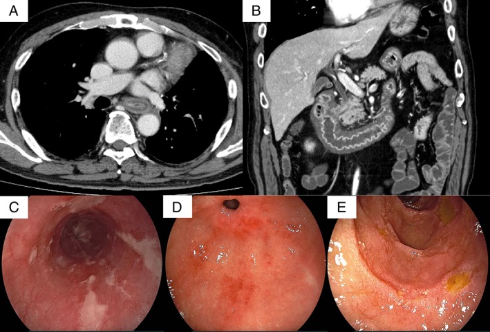
Figure 1.

(A and B) A thoraco‐abdominal contrast CT revealed extensive thickening and oedema in the walls of the oesophagus, stomach, duodenum, and jejunum. (C–E) Oesophageal gastroduodenal endoscopy revealed diffuse longitudinal ulcers of the oesophagus (C), which were different from ulcers observed in cases of reflux esophagitis. The stomach demonstrated erosion in the upper part and mottled reddening near the pyloric ring (D). Mucosal atrophy of the stomach was not found. Small ulcers and flares were scattered in the whole duodenum (D).
