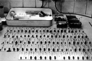
Figure 1.

Crystals of native and heavy metal derivates of myoglobin used for X‐ray analysis. Each of the 100 or so crystals shown in the figure is mounted in a thin‐walled glass tube about 1 mm in diameter. The tubes are sealed at each end to preserve humidity and one end is enclosed in a “lump” of modeling clay that was used to align the crystal prior to X‐ray data collection. Photograph by Bror Strandberg. Courtesy of Bror Strandberg and Medical Research Council Laboratory of Molecular Biology.
