
Figure 8.
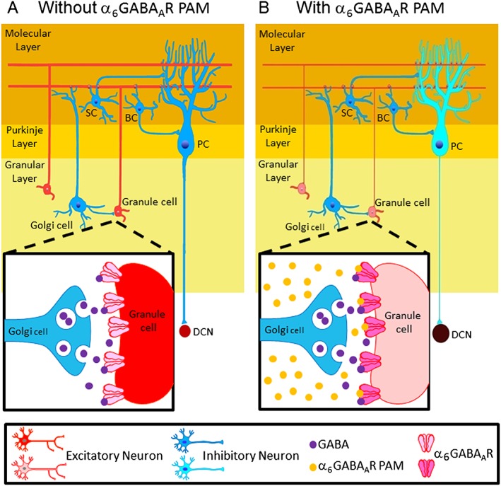
A proposed model for how an α6GABAA receptor PAM affects canonical cerebellar circuits and ultimately leads to the restoration of prepulse inhibition. Schemes of the basic circuit of the cerebellar cortex, which consists of molecular, Purkinje and granule layers, (A) before and (B) after treatment with an α6GABAA receptor PAM, such as hispidulin or Compound 6. The excitatory (red) and inhibitory (blue) neurons in the cerebellar cortex (the yellow part) that may be involved in the action of α6GABAA receptor PAMs, including Golgi cells that form GABAergic synapses onto granule cells, where the α6GABAA receptors are located synaptically and extrasynaptically (enlarged squares). Granule cells form excitatory synapses on dendrites of PCs and also send feedforward inhibition onto PCs indirectly through GABAergic interneurons such as basket cells (BCs) and stellate cells (SCs). PCs are GABAergic output neurons in the cerebellar cortex, providing an inhibitory control onto the downstream DCN. Thinner lines and lighter colours represent reduced neurotransmission and neuronal activity, respectively, after α6GABAA receptor PAM treatment. α6GABAA receptor PAMs act by enhancing Golgi‐GABAergic transmission to granule cells, which then decreases PC activity as a result of the attenuated excitatory inputs from granule cells and this sequence of events ultimately decreases the inhibitory output from the cerebellar cortex. This will activate the DCN, to increase dopamine levels in the PFC, leading to the restoration of prepulse inhibition.