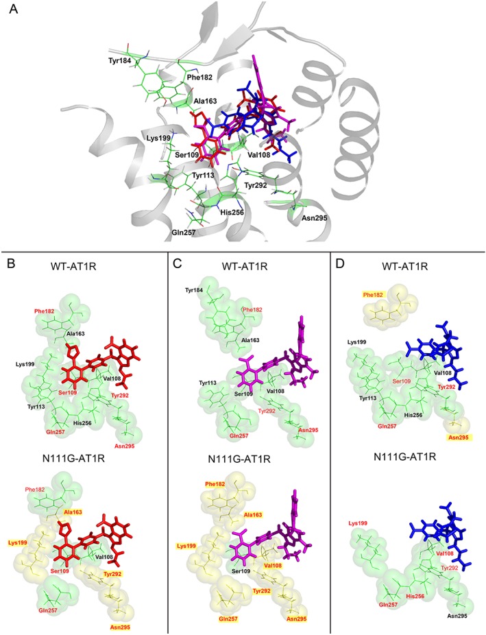
Figure 5.

(A) Molecular model of candesartan (red), telmisartan (magenta) and eprosartan (blue) docking to AT1 receptors. Comparison of the AT1 receptor binding pocket interactions with (B) candesartan, (C) telmisartan and (D) eprosartan in the ground state (WT‐AT1 receptor) and active state (N111G‐AT1 receptor). The ARBs are shown as sticks with red (candesartan), magenta (telmisartan) and blue (eprosartan) carbons. Side‐chain positions of residues investigated in this report are located within a 10 Å pocket for each ARB. In each ARB‐bound model, single side‐chain mutations affecting binding with a >3‐fold change in Ki are indicated by a thick green colour and bold label, both in the ground state (WT‐AT1 receptor) and active state (N111G‐AT1 receptor). A red residue label denotes a significant effect on inverse agonism for IP formation in WT‐ and N111G‐AT1 receptors. Highlighted residues shown as yellow spheres denote a unique influence on inverse agonism for IP formation in the specified state of AT1 receptors for the particular ARB.
