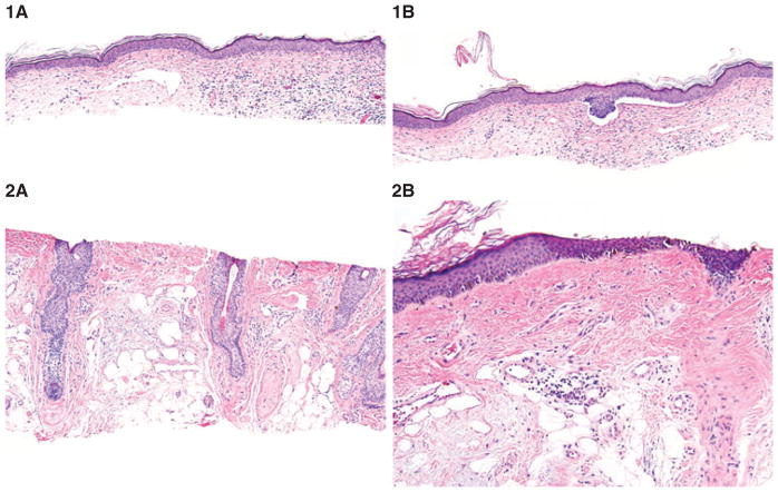
Fig. 6.
Blinded evaluation of the dermal images of stromal changes. 1A) Fibromyxoid dermal changes with interstitial inflammatory infiltrate and widended tortuous vessels are suggestive of basal cell carcinoma; hematoxylin/eosin, ×100. 1B) Further sections reveal superficial basal cell carcinoma; hematoxylin/eosin, ×100. 2A) Dermal changes show red fibrosis pushing the solar elastosis. Although the differentiation between melanoma and actinic keratosis is not unequivocally possible, fibrosis along the hair follicles suggests melanoma in situ; hematoxylin/eosin, ×100. 2B) Further sections reveal irregular single melanocytes and dermal nests, suggestive of melanoma in situ (lentigo maligna type); hematoxylin/eosin, ×100,

