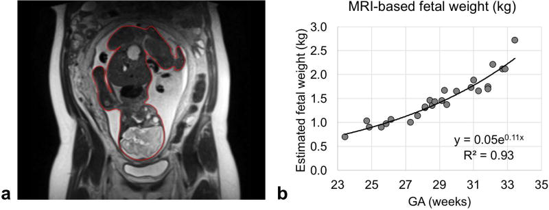
Figure 4.
(a) Representative coronal half-Fourier single-shot turbo spin-echo (HASTE) image of a gravid uterus. Red contour depicts the segmentation of the fetus on a single image. Segmentations of the entire fetal body were used to estimate fetal mass from volume information. Fetal volume was then converted to mass using the relationship determined by Baker et al: mass (kg) = 1.03 · volume (dm3) + 0.12 (kg). (b) Plot of fetal mass in kg versus gestational age (GA).

